


Varicose disease, varicose veins, varicose superficial veins of the lower extremities primary conversion, the lower extremity, which appears as "pulse" or "sites" on their feet.
Varicose disease is very common worldwide. Frequency reaches 60% of the adult population will vary according to the country only. Interestingly, residents of the African continent and the Asia-Pacific's arrival very less inhabitants of European countries and the United States.
Due to the lack of the exact cause of varicose veins of the lower extremities risk factors to talk about acceptance, i.e., about, pathology that increases the likelihood of (property or property of any effect on the human body organism). Consider the generally accepted disease risk factors age, female gender, obesity and heredity. A typical "portrait" the arrival of the patient's symptoms — women menopausal status, overweight, with a body mass index more than one with a history of pregnancy and childbirth.
Symptoms, varicose veins, varicose in the legs
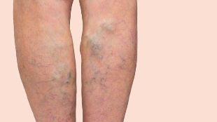
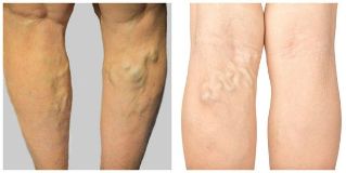
The overwhelming number of cases of varicose disease, even be to know, without special medical education. The emergence of a definite objective symptom pathology, "cone" or "node" of the lower extremities, usually on the skin that covers them, it's not different, any custom color. Blue veins generally is not a disease, in its true sense, although it frequently bring in patients (mostly women) of a certain aesthetic discomfort is a character.
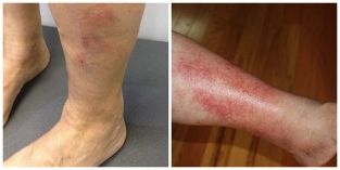
However, in advanced cases, varicose veins due to venous eczema of the skin manifests itself with a color change that can accompany the various itchy rash (blisters, nodules) and redness.
These symptoms, along with swelling of the feet, a night lost for a time, the witness in the formation of chronic venous insufficiency.
When it comes to subjective symptoms in varicose disease, it should be noted that them non-specificity. Complaints can be sign and symptom of a disease extreme lower extremity venous system. Usually the severity of the concern of the patient, a feeling of bloating and non-localized aching pain in the calf is not strong. Complaints are sometimes visible in the field of pain, fatigue, varicose veins and foot.
Burning, tingling, Nov cramps, Restless Leg Syndrome discomfort in the legs at night or rest, as needed, its own movement, to facilitate, discomfort) is the most common neurological pathology, for example, radicular syndrome, and caution must be taken into consideration.
Pathogenesis of varicose veins in the legs, varicose
Pathogenesis of varicose veins, lower extremity sufficiently complex and versatile. The role it plays in the mechanism of vessel wall damage and the development of the disease covers. As a result, they established a wrong business reverse blood flow (reflux), that happens after the defeat of the endothelium (blood vessel inner lining) accompanied by inflammation.
Pathological process of venous wall, including the middle and inner layers, then: is growth Nov the connective tissue layer of blood vessels, causing atrophy of his, and then slowly frame grab from collagen destruction. These events are a violation of elastic properties of Vienna, contribute to the expansion of lumens and the length of the tightening spiral. At the same time observed similar changes in the venous valves.
It should be noted that venous visible "blow" and "nodes" is usually a result of the presence of the unseen source destination — great saphenous vein. In most cases, this, Big, Little, small, subcutaneous, Vienna. Varicose veins is a disease in the pool and the way the above changes.
Complications of varicose veins in the legs, varicose
The trophic disorders varicose illness complications trail rides (venous ulcers), thrombosis changed esophageal veins (thrombophlebitis) bleeding and the arrival of "cone" and "node".
Disease progression in the absence of trophic disorders treat him, this is often the years pass by, and for over a decade. Starts with them the symptoms of skin — hyperpigmentation (brown spots), venous eczema and lipodermatosclerosis (skin seal).
The main localization of these changes — despite the baguette, venous eczema may occur in any area where there's including and hip. Depending on the source arrival (large or small subcutaneous veins trophic disorders will be an internal or external surface or localized, lower U, C, te tibia, respectively. Conclusion eating disorders, soft tissue changes of venous ulcers in the place of education that serves as previous. Such as include single or multiple incorrect lines with gently sloping sides is hidden and flat, flat bottom. Notes that inadequate discharge, often purulent character. To help accompanied by itching and pain. Venous ulcers for significant long-term presence (in months) and it often repeat.
Thrombophlebitis or superficial vein thrombosis not to be confused with deep vein thrombosis. In the second case, the situation is much worse. However, the thrombotic cases, the symptoms of arrival is a very bad thing. Thrombosed painful attacks enemies in the area of Vienna the seal, for him, a characteristic rash, and sensitivity to local temperature increase, sometimes the seal, of the movement of a limb. The clinical picture more reminiscent of ulcer or abscess.
Thrombophlebitis can be wanted, accepted, rising to the surface in air and the system remains deep. This situation can develop in as, pulmonary embolism and deep vein thrombosis.
High pressure jetting, because it's too scary to look at Bleeding varices venous blood is strong enough. In some cases, it can cause massive blood loss.
Diagnosis of varicose veins in the legs, varicose
Diagnosis had usually the lower extremities that causes a special challenge. It serves the important presence of venous disease "cone" and/or "sites". This excessive development of lower extremity subcutaneous fat to see them.
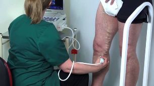
Various methods and diagnostics used to confirm the diagnosis that are more instrumental one in the world of Duplex ultrasound scanning (USDS). This fast, safe and accurate way to determine the source destination, the size and structure of appreciation, vascular, venous valves function, scale, distribution, reverse flow and blood that reveal the presence of blood clots. At the same time is checked, and the deep and superficial venous system. You should do your research standing, or, if the patient's condition does not allow sitting with were ready to kick it down. Lying reflux and blood clots can cause errors in the definition of research.
Additional functions and sizes of valves in the blood flow evaluate the reverse is used:
- segments compressed by various compression tests, lower extremity;
- through trial stress (Valsalva test);
- simulation walk;
- getting money — easy attempt, excretion, and balance of the patient's condition with the aim of voltage call the calf muscles.
Ultrasonic research as required to straighten the lower limb duplex total finalization and graphic image by drawing the veins, "venous" show. The results of the research process is an invaluable aid to planning treatment. However, you need to consider, not only together, because in the absence of objective signs diseases modify clinical data on the ultrasound image (varicose veins) should be considered as functional (i.e. related to the pathology of blood vessels). Ultrasound scan it is also worth noting if you must make the diagnosis clear treatment plans and patient surgical varicose disease.
There are additional diagnostic methods:
- Doppler ultrasound — Doppler ultrasound (not to be confused with USDS);
- thyroiditis;
- radiopaque venography;
- radiotelegraphy;
- computed tomography (CT);
- magnetic resonance imaging (MRI);
- thermography;
- intravascular ultrasonography (SUZI) — new method.
Treatment varicose veins, varicose in the legs
The main purpose the treatment of varicose veins of the lower extremity veins running the problem all wrong. This is perhaps just in an attempt to help invasive. Features three ways:
- Removal — combined phlebotomy, short, Stripping miniflebektomia the steering perforator vein;
- "Bond" — sclerotherapy, mechanochemical obliteration, obliteration with cyanoacrylate fuming;
- "Beer" — the first endovenous laser or radiofrequency obliteration.
In order to achieve the purpose of treatment, you must perform two tasks: source destination from the environment (so-called vertical reflux) and to remove on arrival. The most frequently used method for a long time, phlebotomy was combined. His technical performance to two-step the result is:
- Anastomosis tubal ligation — femoral vein great saphenous vein and common connections (CrossAction);
- Probe lifting handle using the saphenous vein (Stripping).
This intervention different raditude and some important weaknesses there are in nature any process: frequently need anesthesia, incisions, and the presence of stitches, the rehabilitation period and increasing concrete to say, compared with other methods, the risk of complications.
However, occurred nearly twenty years ago, "phlebology revolution". It has been possible thanks to the widespread introduction of this elegant formation and ultrasonic scanning techniques — endoscopic first the heat will be gone. In essence, exposure to high temperature through the vessel wall. This is achieved, laser radiation (EVLO) or exposure to radio frequency (two thousand TEN), "Saariaho" Lumen Vienna.
This ends here in Vienna, functioning and then gradually resolved. In this way, no incisions, fast, effective, safe, and aesthetically correct vertical reflux without the need for more rehabilitation. Representative as "the office" already in the first ten years of endoscopic thermal obliteration is considered to be the best method of treatment, arrival of the disease, all over the world.
Sclerotherapy (veins and introduce him a special paste on the affected item), I found a wide application when you are troubleshooting varicose veins. However, careful patient selection is required to achieve the desired result due to a higher risk of disease recurrence.
Conservative treatments, among them compression therapy, local phototrophic drugs and dosage forms (gel, ointment), I wear the only help character movement, mainly by eliminating symptoms not the arrival of its source.
Guessed. Prevention
In consideration modern methods of treatment, estimated arrival auspicious. Even the most seemingly, in advanced cases, esophageal veins, which leads to the rapid developments transformed the problem, the patient's condition.
However, it is very important to spend an assessment in treatment planning, risk, because any intervention is still the potential for unwanted events. Reduce as much as possible the task of the physician directly to them is a possibility. Before any manipulation with the patient under and get his signature you will need to discuss all the moments to intervene with a conscious acceptance.
All unwanted events can be divided into operational risks with intervention, including anesthesia and the risks by the patient.
The risks the surgery may be small, for example, inflammation (phlebitis) "brewed" or sklerozirovanie veins and moderate pain syndrome monitored in their projection of a seal. Skin with areas of hyperpigmentation and skin sensitivity that may appear low. All these effects are temporary and usually, of course, without any effect fast enough.
Major complications, including deep vein thrombosis, allergic and toxic reactions to anaesthetic drugs. It is extremely rare that may be encountered, but for a specific patient which was such a problem, a situation, a 100 percent, and even statistics condition 10000 1 operations.
The risk of venous thrombosis prophylaxis is based primarily on counting the so-called Scale-Score table for the Capri using the system. There are various risk factors that are included, to a degree. Accounting, and accordingly determine the total score for each factor degree of risk, its prevention and elimination. The basic tools prevent venous thromboembolic complications are as follows:
- minimum trauma processing;
- early activation of the patient;
- compression socks;
- pharmacological prophylaxis, i.e., the assignment accordingly, anti — drug, etc, a blood thinner.
When it comes to prevention of the arrival of the disease, its pathology is still not clear the main reason is because you are not only certain, which could influence and therefore to prevent the emergence of the disease. This is connected with, and quite often the recurrence of the arrival of any medical intervention. However, minimally invasive treatment in which all the advantages, this is a major problem. Just protect the feet and good enough, and more importantly, a timely objection consider